Research
Microscopy
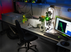
PerkinElmer UltraVIEW ERS Spinning Disk Confocal Imager
Confocal microscopy is an optical imaging technique used to increase optical resolution and contrast of a micrograph by using point illumination and a spatial pinhole to eliminate out-of-focus light in specimens that are thicker than the focal plane. It enables the reconstruction of three-dimensional structures from the obtained images.
Our imager includes the PerkinElmer UltraVIEW ERS spinning disk confocal imager with Krypton 561 and Argon 488/515 gas lasers and solid-state lasers emitting at 405, 440, 515, and 640nm. Emission discrimination includes the following filters:
- 527(W55)
- 445(W60), 615(W70)
- 485(W60), 705(W90)
- 587(W125)
The imaging system utilizes a Zeiss Axiovert 200 inverted microscope equipped with a piezo Z-Focus drive for 3-D imaging. Additionally a Solent brand environmental chamber for live cell imaging is available. Our confocal unit utilizes the following Zeiss objectives:
- 20x/0.8 Plan-Apochromatic 0.55/0.17
- 40x/1.3 Oil Fluar 0.16/0.17
- 40x/0.4 LD Plan-Neofluar 2.0/2.0
- 63x/1.4 Oil DIC Plan-Apochromat 0.19/0.17
- 100x/1.4 Plan Apochromat 0.17/0.17
Images are acquired and analyzed using Volocity software on a Dell PC. Volocity enables the use of automated capture protocols, 3D reconstruction, FRET, colocalization analysis and 3D ratio imaging.
Tutorials:
- Importing and Exporting Data in Volocity
- Performing Simple Measurements in Volocity
- Colocalization
- Tracking
- 3D Visualization and Movie Making in Volocity
- Creating Measurement Protocols
- Deconvolution in Volocity
As with all forms of microscopy sample preparation is key. To be compatible with laser imaging and our objectives working distances samples should be prepared properly and guarded by a 0.17mm (No.1) cover slip against a glass slide.
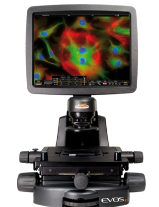
AMG EVOS fl LED Fluorescent Microscope
- Easy to use inverted multichannel LED fluorescence microscope
- Screen slides - DAPI, Green and Red fluorescence
- Phase contrast transmitted light
- Check transfection - easy tools for overlay and counting
- Specimen holders are available for slides, multi-well plates, 100mm and 35mm dishes, and various flasks.
- LED illumination - no Arc lamp warm up
- High resolution monochrome camera
- Simple, easy-to-use software
- Save images direct to USB drive
Advanced Microscopy Group (AMG) EVOS FL website
Detailed specifications:
Objectives:
- 4X/0.13 LWD Ph/Fl (works with plastic culture dishes, image through slide rather than coverslip for slides)
- 10X/0.25 LWD Ph/Fl (works with plastic culture dishes, image through slide rather than coverslip for slides)
- 20X/0.40 LWD Ph/Fl (works with plastic culture dishes, image through slide rather than coverslip for slides)
- 40X/0.75 FL corrected for coverglass number 1.5, 0.17 mm
Imaging channels:
- Phase contrast transmitted light imaging
Fluorescent LED light cubes with fully variable intensity control:
- DAPI (357/44, 447/60)
- GFP (470/22, 525/50)
- TxRed (585/30, 624/40)
Stage and sample capabilities:
- Manual stage with stage adapters for most commonly used sample vessels
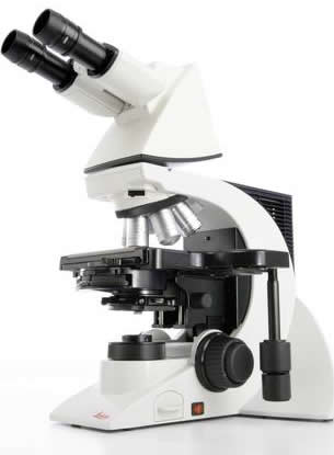
Leica DM2000 Upright Fluorescent Microscope
Leica DM2000 Upright Fluorescent Microscope with the following noteworthy specifications:
- 5x, 10x, 20x, 40x & 100x Brightness synchronizers objective lenses
- Blue, Green, Red & BGR filter cubes
- Spot Software - Basic
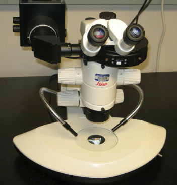
Leica MZ6 Dissecting Fluorescent Microscope
Leica DM2000 Upright Fluorescent Microscope with the following noteworthy specifications:
- 0.1-10x magnification
- FITC filter and mercury lamp
- SPOT imaging software
| Microscopy Service | Description | Dissecting Fluorescent Leica MZ6 | Upright Fluorescent Leica 2000 | Confocal Ultraview ERS, Axiovert II, Volocity Software |
|---|---|---|---|---|
| Acquisition | Core facility technician will capture images. Does not include sample preparation. | $29.00 / hour | $29.00 / hour | $34.00 / hour |
| Assisted | Core facility technician will assist user with capturing images. No training is required. | $32.00 / hour | $32.00 / hour | $40.00 / hour |
| Unassisted | Fee for usage of this equipment. ALL USERS MUST COMPLETE TRAINING | $8.00 / hour | $8.00 / hour | $21.00 / hour |