In the Electron Microscopy lab, the girls learned how to use both of the most powerful, expensive microscopes available at the College of Marine Science: the transmission electron microscope, or TEM, and scanning electron microscope, or SEM.
The TEM passes a focused beam of electron through a specimen, allowing one to view materials on the order of nano meters. With the TEM, the girls were able to look into the middle of cells, and see all the organelles, including the mitrochondria, the “powerhouse” of a cell. They were also able to see bacteria, and even individual viruses.
The beam of the SEM, rather than passing through the specimen, creates a raster pattern reflected off a the specimen’s surface, allowing the creation of a two- or three-dimensional image.
The girls towed a bottle off the sea wall to catch plankton; they also prowled campus,
capturing insects and other tiny specimens to image under the SEM.
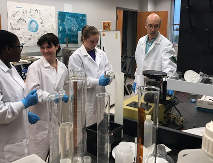
They then placed the samples in chemical preservative, sputter-coated them with a
gold-palladium alloy, placed them in the vacuum chamber, and learned to “drive” the
enormous scanning electron microscope, saving their images for the last day of camp.